Category Archives: internal medicine

Genome-wide screen of learning in zebrafish identifies enzyme important in neural circuit

Cardiology_Internal Medicine_NeurologyImplications for understanding underlying molecular genetics of human neuropsychiatric disorders, according to Penn study

Researchers at the Perelman School of Medicine at the University of Pennsylvania describe the first set of genes important in learning in a zebrafish model in the journalNeuron this week. “Using an in-depth analysis of one of these genes, we have already revealed an important relevant signaling pathway,” says senior author Michael Granato, PhD, a professor of Cell and Developmental Biology. “The proteins in this pathway could provide new insights into the development of novel pharmacological targets.”Over the last 20 years, zebrafish have become great models for studying development and disease. Like humans, zebrafish are vertebrates and over 80 percent of human genes bearing disease descriptions are also present in zebrafish. As such, this animal model has become increasingly popular to study human diseases such as cardiovascular conditions or tumor formation.

Studies show young people ‘wish they were better informed about sex’

Pediatrics_Internal Medicine_General Practice_OBGYN_Family MedicineResearchers investigating how young men and women learn about sex found that a gap exists between the type of sex education young people want and what they receive.The researchers also identified differences between how men and women learn about sex and relationships. The two studies, published in BMJ Open, utilized data from the third National Survey of Sexual Attitudes and Lifestyles (Natsal-3) – the largest scientific study of its kind in the UK.”The terrain young people have to navigate as they are growing up has changed considerably over the past 20 years and it will inevitably continue to do so,” says study author Dr. Clare Tanton. “This means that while we need a more structured approach towards sex and relationships education, we must also be able to adapt to these changing needs.”In one of the two studies, data from Natsal-3 for 3,869 participants, conducted between 2010 and 2012, was compared with data from surveys conducted in 1990-1991 and 1999-2001, in order to assess how sources of information about sex may have changed over time.
Read the rest of the article at http://www.medicalnewstoday.com/articles/290528.php.

One in 4 patients who visited emergency department for chest pain did not receive follow-up care

Cardiology_IM_FM_GP_Emergency MedicinePatients with multiple health issues and who are at higher risk of adverse events are less likely to receive follow-up care from a physician after visiting an emergency department for chest pain, reports a study published in CMAJ(Canadian Medical Association Journal).Chest pain is one of the most common reasons people visit emergency departments, with about 500 000 visits every year in Canada alone.The study looked at 56 767 patients with chest pain who visited an emergency department in Ontario between April 2004 and March 2010. Of these, 42 535 (75%) were seen by a primary care physician or cardiologist within 30 days after discharge, and 14 232 (25%) did not receive follow-up care. Patients with multiple illnesses such as heart disease, kidney disease and dementia, and rural residency were less likely than others to receive follow-up care in the following month. Patients who had visited a primary care physician or cardiologist in the preceding year were more likely to see a physician after discharge.”We initially thought that emergency department physicians would risk stratify patients such that those with more medical and cardiac comorbidities would receive more physician follow-up care,” writes Dr. Dennis Ko, a senior scientist at the Institute for Clinical Evaluative Sciences (ICES) and an interventional cardiologist at the Schulich Heart Centre, Sunnybrook Health Sciences Centre, Toronto, Ontario, with coauthors. “However, patients with more medical comorbidities in our cohort were less likely to receive physician follow-up postdischarge.”

Read the rest of the article at  http://www.medicalnewstoday.com/releases/289784.php.

Novel peptide shows promise in penetrating heart attack scar tissue to regenerate cardiac nerves and avert dangerous arrhythmias

Cardiology_IM_GP_FMCase Western Reserve’s chemical compound aimed at restoring spinal cord function may have an additional purpose: stopping potentially fatal arrhythmias after heart attack.Case Western Reserve neurosciences professor Jerry Silver, PhD, long has believed that lessons learned over decades from spinal cord research could someday apply to other areas of the body. He got the chance to test his theory when a colleague from another campus realized that his new compound – intracellular sigma peptide (ISP) – could address a critical cardiac issue.The results of the project, led by Oregon Health & Sciences University (OHSU) researcher Beth A. Habecker, PhD, exceeded even Silver’s greatest hopes: 100 percent success in animal models. Details can be found in Nature Communication.”Essentially, the OHSU group cured arrhythmia in the mouse using ISP,” Silver said. “They observed true regeneration right back into the scar within the infarct area. This is pretty exciting.”

Read the rest of the article at  http://www.medicalnewstoday.com/releases/289007.php.

Early detection of heart attacks aided by gold nanoparticles

Cardiology_IM_FM_GP-Oncology_Dentistry_Hematology

NYU Polytechnic School of Engineering professors have been collaborating with researchers from Peking University on a new test strip that is demonstrating great potential for the early detection of certain heart attacks.

Kurt H. Becker, a professor in the Department of Applied Physics and the Department of Mechanical and Aerospace Engineering, and WeiDong Zhu, a research associate professor in the Department of Mechanical and Aerospace Engineering, are helping develop a new colloidal gold test strip for cardiac troponin I (cTn-I) detection. The new strip uses microplasma-generated gold nanoparticles (AuNPs) and shows much higher detection sensitivity than conventional test strips. The new cTn-I test is based on the specific immune-chemical reactions between antigen and antibody on immunochromatographic test strips using AuNPs.

Compared to AuNPs produced by traditional chemical methods, the surfaces of the gold nanoparticles generated by the microplasma-induced liquid chemical process attract more antibodies, which results in significantly higher detection sensitivity.

cTn-I is a specific marker for myocardial infarction. The cTn-I level in patients experiencing myocardial infarction is several thousand times higher than in healthy people. The early detection of cTn-I is therefore a key factor of heart attack diagnosis and therapy.

The use of microplasmas to generate AuNP is yet another application of the microplasma technology developed by Becker and Zhu. Microplasmas have been used successfully in dental applications (improved bonding, tooth whitening, root canal disinfection), biological decontamination (inactivation of microorganisms and biofilms), and disinfection and preservation of fresh fruits and vegetables.

The microplasma-assisted synthesis of AuNPs has great potential for other biomedical and therapeutic applications such as tumor detection, cancer imaging, drug delivery, and treatment of degenerative diseases such as Alzheimer’s.

The routine use of gold nanoparticles in therapy and disease detection in patients is still years away: longer for therapeutic applications and shorter for biosensors. The biggest hurdle to overcome is the fact that the synthesis of monodisperse, size-controlled gold nanoparticles, even using microplasmas, is still a costly, time-consuming, and labor-intensive process, which limits their use currently to small-scale clinical studies, Becker explained.

Adapted by MNT from original media release

http://www.medicalnewstoday.com/releases/288143.php


Researchers develop a new distance rehabilitation system for patients with heart pathologies

Cardiology_Internal Medicine_General Practice_Family Medicine

A team of Spanish researchers of the Universitat Politècnica de València, the Universidad Politécnica de Madrid and the company Medtronic have participated in the development of a new distance heart rehabilitation system based on physical exercise routines for people affected by heart pathologies.

The system is designed for both chronic patients and the recovery of people who have suffered a heart event (for instance, a heart attack) or if they have had heart surgery. In any of these cases, it helps patients to exercise and adopt a healthy lifestyle.

HeartCycle GEx has been developed within the European project HeartCycle. This system creates an online connection between the cardiologist in the hospital and the patients in their homes. The latter, equipped with sensorized t-shirts, do the rehabilitation exercises while their mobile telephones receive all the information about their heart and respiration rate, and messages to make more or less effort according to the doctor’s instructions, etc.

Its main advantage is its ability to motivate patients; GEx helps patients follow the rehabilitation program in an easier and more entertaining way. To that end, GEx incorporates multimedia content, an avatar, as well as graphic information about the indicators related to the patient’s performance (heart rate, effort level, etc).

“The Achilles’ heel of heart rehabilitation is that patients abandon it in a few weeks, so its effect is watered down. If the user is motivated, the level of compliance will increase and their health will improve. The most important aspect is to improve adherence to the rehabilitation plan and maintain it over time, and this system does just that”, explains Alvaro Martínez, researcher at the ITACA Institute of the Universitat Politècnica de València.

In addition, GEx will be connected to the hospital information systems in order to guarantee optimum, personalised health care for the patient. The system enables each user’s condition to be monitored and the response to the instructed therapy assessed, so personalised plans can be created and any deterioration in health status that requires immediate intervention can be detected.

The doctor has a web application in which to program the personalised rehabilitation plan that is sent to the patient – the prescribed plan is adapted to the patient’s physical condition, who performs an effort test – to design the most suitable prescription possible.

“Every time the patient does one of the exercises, the system sends new data, so the doctor can immediately determine whether the patient’s effort meets the standard set and make adjustments to the prescription if necessary”, adds Alvaro Martínez.

This doctor’s web application was entirely developed at the ITACA Institute of the Universitat Politècnica de València.

“Today, the heart rehabilitation programs in Spain take place in hospitals or specialized centres. HeartCycle GEx is a different and convenient alternative to access to those programs since the patients can follow them anywhere, even at home, maintaining the level of health care that they receive”, says Cecilia Vera, researcher at the Life Supporting Technologies Group of the Universidad Politécnica de Madrid.

Validation in hospitals

GEx has been validated with 132 patients at three hospitals in Spain, Germany and the United Kingdom and, according to the tests performed, it provides better results than traditional rehabilitation programs, “This improvement is seen in the patient’s functional capacity, as well as in weight loss and a reduction in cholesterol levels”, says Alvaro Martínez.

“HeartCycle GEx is an initial step to the new heart rehabilitation systems of the future, providing personalised solutions, adapted to each patient and accessible from any location”, adds Cecilia Vera.

The results have been released in the European Journal of Preventive Cardiology.

Adapted by MNT from original media release

Picture courtesy of medicalxpress.com.

http://www.medicalnewstoday.com/releases/287370.php


High fitness levels reduce hypertension risk

Cardiology_IM_Nurses_GP_FM

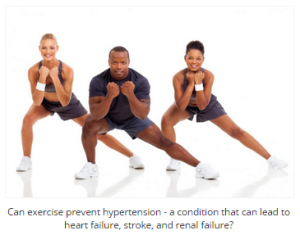
While being physically fit is beneficial in and of itself, researchers now report that people with high levels of fitness are less likely to develop high blood pressure – also referred to as hypertension – a risk factor for cardiovascular disease.

The study, published in the Journal of the American Heart Association, examined the association of fitness with hypertension among participants undergoing treadmill stress tests to rule out ischemia as a cause of chest pain or shortness of breath.

“If you’re exercising and you’re fit, your chances of developing hypertension are much less than someone else who has the same characteristics but isn’t fit,” says Dr. Mouaz H. Al-Mallah, senior author of the study.

Normal blood pressure is below 120/80 mm Hg – the first number (systolic measurement) represents peak pressure in the arteries and the second number (diastolic measurement) represents minimum pressure in the arteries. Blood pressure is considered to be high when it is greater than 140/90 mm Hg.

There are two types of hypertension. While secondary hypertension appears suddenly and is caused by underlying conditions such as kidney or thyroid problems, primary hypertension has no identifiable cause and develops gradually over the course of many years.

In the US, hypertension affects 1 in 3 adults. According to the American Heart Association (AHA), 78 million people in the country have been diagnosed with the condition.

“Hypertension is associated with a lot of other illnesses and adds significantly to health care costs,” explains Dr. Al-Mallah, “so we need to know how we can reduce it.”

Measuring physical fitness and high blood pressure

The researchers assessed 57,284 participants from the Henry Ford Exercise Testing (FIT) Project, from 1991-2009, taking treadmill stress tests. Of these, 35,175 participants had a history of hypertension.

The team measured the physical fitness of the participants by estimating how much oxygen their bodies used per kg ofbody weight per minute, and thus how much energy they burned in metabolic equivalents (METs).

With 1 MET representing the amount of energy expended by the body at rest, a large number of METs reflects a high-intensity workout.

The researchers observed that participants whose most intense exercise was less than 6 METs had more than a 70% likelihood of having hypertension at the start of the study. Conversely, participants whose maximal exercise output was 12 METs were less than 50% likely to have hypertension.

During the stress test, participants who managed to reach 12 METs or more were 20% less likely to develop hypertension compared with participants who reached less than 6 METs.

A total of 8,053 new cases of hypertension were reported in participants’ medical records and administrative claims during the study’s follow-up period. Of these new cases, 49% were among participants with the lowest fitness (less than 6 METs), and only 21% were among participants with the highest fitness (more than 12 METs).

Fitness: a ‘strong predictor’ of hypertension

Although the study uses a large and diverse population sample, the participants were all originally referred for a stress test, indicating that their initial cardiovascular disease risk would be greater than that of the general population, potentially hindering the generalizability of the findings. The study was also limited by a lack of measuring incidental hypertension in a clinical setting.

Dr. Al-Mallah states that further study is required in order to determine how increasing and decreasing fitness levels affect the risk of hypertension over time. Physical activity was not formally assessed in the study, and this could be addressed in future research as well.

Hypertension is a major risk factor for cardiovascular disease, the number one cause of premature mortality in the developed world. High levels of exercise have been associated with protecting the body from certain health conditions, and now this study suggests adding hypertension to the list.

“Fitness is a strong predictor of who develops hypertension and who does not,” says Dr. Al-Mallah. “This is a clear message to everyone: patients, physicians and lawmakers. It’s very important to be fit.”

Medical News Today also recently reported on a study suggesting that sugars may contribute more to hypertension risk than salt.

Written by James McIntosh

http://www.medicalnewstoday.com/articles/287109.php


‘Obesity can reduce life by up to 8 years’

Cardiology_IM_FM_GP

Life expectancy can be reduced by up to 8 years by obesity, which can also cause adults to lose as much as 19 years of healthy life if it leads to type 2 diabetes and cardiovascular disease. A study published in The Lancet Diabetes & Endocrinology examines the issue.


The researchers behind the study analyzed data from the US National Health and Nutrition Examination Survey (NHANES), creating a disease-simulation model to estimate the risk of adults of different body weightdeveloping diabetes and cardiovascular disease.

From this, the researchers then calculated the extent to which overweight and obesity may contribute to both years of life lost and years of healthy life lost in American adults aged between 20 and 79 years old, in comparison to people of normal weight.

They found that people who were overweight (BMI 25-30 kg/m2) were estimated to lose up to 3 years of life, depending on age and gender. Individuals classed as obese (BMI 30-35 kg/m2) were calculated to lose up to 6 years, and people classed as very obese (BMI 35 kg/m2 or more) could lose up to 8 years of life.

According to the study, excess weight had the greatest impact on lost years of life among the young and dropped with increasing age.

Obesity can cause the loss of up to 19 ‘healthy life-years’

As well as reducing life expectancy, carrying extra weight was also found to reduce “healthy life-years,” which were defined in the study as years free of obesity-linked cardiovascular disease and diabetes.

Young adults aged between 20 and 29 showed the highest losses of healthy life-years, adding up to around 19 lost years for very obese people. Among people who were overweight or obese, the researchers calculated that two to four times as many healthy life-years were lost than total years of life lost.

Dr. Steven Grover, lead author and professor of medicine at McGill University and a clinical epidemiologist at the Research Institute of the McGill University Health Centre in Canada, explains the findings:

“The pattern is clear. The more an individual weighs and the younger their age, the greater the effect on their health, as they have many years ahead of them during which the increased health risks associated with obesity can negatively impact their lives.

These clinically meaningful calculations should prove useful for obese individuals and health professionals to better appreciate the scale of the problem and the substantial benefits of a healthier lifestyle, including changes to diet and regular physical activity.”

This week on Medical News Today, we also looked at a study published in The BMJ that found obesity during early pregnancy is a risk factor for infant mortality.

The researchers behind that study found that infant mortality was “moderately increased” among overweight and mildly obese mothers (BMI 25-35 kg/m2) compared with mothers of a normal weight; but among more obese mothers (BMI over 35 kg/m2), the risk of infant mortality was more than doubled.

We also reported on a study in the journal Preventing Chronic Disease that found women – particularly black women – are more at risk of increased obesity if they work jobs that involve a lot of sitting down.

Written by David McNamee

http://www.medicalnewstoday.com/articles/286518.php


Scientists identify a rise in life-threatening heart infection

Cardiology_Dentistry_Internal Medicine_Infectious Disease

Scientists at the University of Sheffield have identified a significant rise in the number of people diagnosed with a serious heart infection alongside a large fall in the prescribing of antibiotic prophylaxis to dental patients.

The pioneering study is the largest and most comprehensive to be conducted with regards to the National Institute for Health and Care Excellence (NICE) guidelines, which recommended dentists should no longer give antibiotics before invasive treatments to people considered at risk of the life threatening heart infection, Infective Endocarditis (IE), which in 40 per cent of cases is caused by bacteria from the mouth.

The team of international researchers, led by Professor Martin Thornhill at the University of Sheffield’s School of Clinical Dentistry, discovered that since the NICE guidelines were introduced in March 2008, there has been an increase in cases of Infective Endocarditis above the expected trend. By March 2013 this accounted for an extra 35 cases per month.

They also identified that the prescribing of antibiotic prophylaxis fell by 89 per cent from 10,900 prescriptions a month, before the 2008 guidelines, to 1,235 a month by March 2008.

Martin Thornhill, Professor of Translational Research in Dentistry at the University of Sheffield, said: “Infective Endocarditis is a rare but serious infection of the heart lining. We hope that our data will provide the information that guideline committees need to re-evaluate the benefits, or not, of giving antibiotic prophylaxis.

Professor Thornhill stressed that healthcare professionals and patients should wait for the guideline committees to evaluate the evidence and give their advice before changing their current practice.

He added: “In the meantime, healthcare professionals and patients should focus on maintaining high standards of oral hygiene. This will reduce the number of bacteria in the mouth which have the potential to cause Infective Endocarditis and reduce the need for invasive dental procedures to be performed.”

The data analysed by an international collaboration of experts from the University of Sheffield, Oxford University Hospitals NHS Trust, Taunton and Somerset NHS Trust, and the University of Surrey in the UK, as well as from the Mayo Clinic and the Carolinas HealthCare System’s Carolinas Medical Center in the USA, is published in The Lancet and will be presented to more than 19,000 delegates from across the world at the American Heart Association annual meeting in Chicago.

The research was funded by a grant from national heart charity Heart Research UK, healthcare provider Simplyhealth and the National Institute for Dental and Craniofacial Research (NIDCR).

Barbara Harpham, National Director of Heart Research UK, said: “The findings play an important part in the ongoing exploration of the link between dental and heart health.

“Projects such as this one are vital to the ongoing collation of evidence to support our understanding of how oral health can impact upon the heart and other conditions within the body. We are committed to furthering medical research in the UK and welcome these new findings.”

http://www.medicalnewstoday.com/releases/285754.php


The heart’s own immune cells can help it heal

Cardiology_Rheumatology_IM_GP_FM

The heart holds its own pool of immune cells capable of helping it heal after injury, according to new research in mice at Washington University School of Medicine in St. Louis.

Most of the time when the heart is injured, these beneficial immune cells are supplanted by immune cells from the bone marrow, which are spurred to converge in the heart and cause inflammation that leads to further damage. In both cases, these immune cells are called macrophages, whether they reside in the heart or arrive from the bone marrow. Although they share a name, where they originate appears to determine whether they are helpful are harmful to an injured heart.

In a mouse model of heart failure, the researchers showed that blocking the bone marrow’s macrophages from entering the heart protects the organ’s beneficial pool of macrophages, allowing them to remain in the heart, where they promote regeneration and recovery. The findings may have implications for treating heart failure in humans.

The study is now available in The Proceedings of the National Academy of Sciences Early Edition.

“Researchers have known for a long time that the neonatal mouse heart can recover well from injury, and in some cases can even regenerate,” said first author Kory J. Lavine, MD, PhD, instructor in medicine. “If you cut off the lower tip of the neonatal mouse heart, it can grow back. But if you do the same thing to an adult mouse heart, it forms scar tissue.”

This disparity in healing capacity was long a mystery because the same immune cells appeared responsible for both repair and damage. Until recently, it was impossible to distinguish the helpful macrophages that reside in the heart from the harmful ones that arrive from the bone marrow.

The new research and past work by the same group – led by Douglas L. Mann, MD, the Tobias and Hortense Lewin Professor of Medicine and cardiologist-in-chief at Barnes-Jewish Hospital – appear to implicate these immune cells of different origins as responsible for the difference in healing capacity seen in neonatal and adult hearts, at least in mice.

“The same macrophages that promote healing after injury in the neonatal heart also are present in the adult heart, but they seem to go away with injury,” Lavine said. “This may explain why the young heart can recover while the adult heart can’t.”

Because they are interested in human heart failure, Lavine and his colleagues developed a method to progressively damage mouse cardiac tissue in a way that mimicked heart failure. They compared the immune response to cardiac damage in neonatal and adult mouse hearts.

The investigators found that the helpful macrophages originate in the embryonic heart and harmful macrophages originate in the bone marrow and could be distinguished by whether they express a protein on their surface called CCR2. Macrophages without CCR2 originate in the heart; those with CCR2 come from the bone marrow, the research showed.

Lavine and his colleagues asked whether a compound that inhibits the CCR2 protein would block the bone marrow’s macrophages from entering the heart.

“When we did that, we found that the macrophages from the bone marrow did not come in,” Lavine said. “And the macrophages native to the heart remained. We saw reduced inflammation in these injured adult hearts, less oxidative damage and improved repair. We also saw new blood vessel growth. By blocking the CCR2 signaling, we were able to keep the resident macrophages around and promote repair.”

Some CCR2 inhibitors are being tested in phase 1 and 2 clinical trials for treating rheumatoid arthritis. But before these drugs can be evaluated in people with heart failure, more work must be done to find out whether the same mechanisms are at work in human hearts, according to the researchers.

“We have identified similar immune cell subtypes that are present in the human heart,” Lavine said. “We need to find out more about their roles in heart failure in patients and understand more about how macrophages that reside in the heart promote repair.”

http://www.medicalnewstoday.com/releases/284750.php